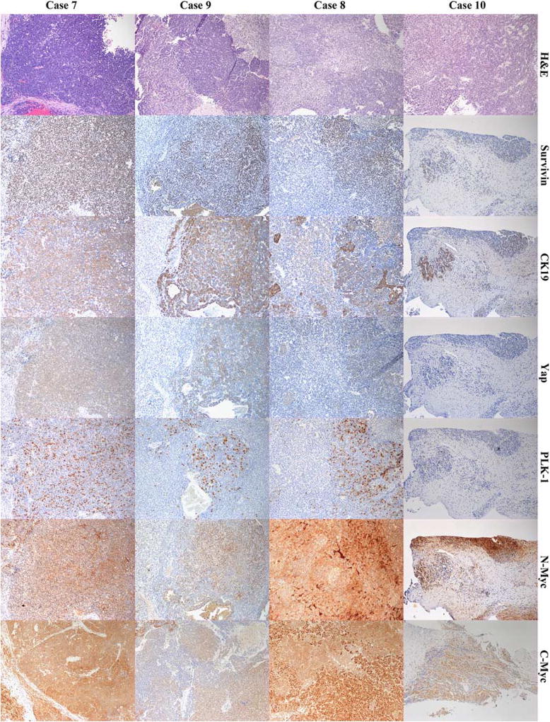
FIGURE 1.
Composite image of 4 cases of hepatoblastoma, fetal-embryonal epithelial type. First row labeled as H&E shows hematoxylin and eosin-stained sections of aggressive tumors (case 7 and 9) and good clinical outcome tumors (case 8 and 10). Shown in the rows accordingly labeled are immunohistochemical stains for Survivin, CK19, Yap, PLK-1, N-Myc, and C-Myc for the 4 cases. All images have a final magnification of × 100.

