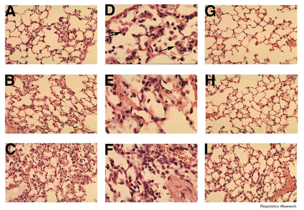
Figure 1.

Pulmonary histopathology during pneumovirus infection. Lungs from mice infected with PVM (60 pfu) for (A, D) 3 days, (B, E) 5 days, and (C, F) 7 days. Images at lower magnification (A, B, and C × 400) show the extent of inflammatory changes, whereas the higher power images (D, E, and F ×1000) demonstrate that the cells are predominantly granulocytes. The arrows in panel D highlight the presence of eosinophils. Lung histology from animals infected with RSV (1 million pfu) for (G) 3, (H) 5, and (I) 7 days (× 400). Note the paucity of acute inflammatory changes.
