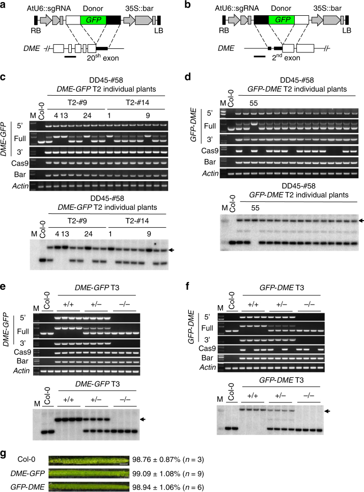
Fig. 3.
GFP knock-in into the endogenous DME locus by gene targeting. a, b Schematics showing HDR donor transgene constructs and part of the targeted DME locus for DME-GFP and GFP-DME knock-in, respectively. The horizontal lines indicate the positions of probes for Southern blotting. c−f Genotyping PCR and Southern blotting for individual T2 lines (b) and T3 plants (e) of DME-GFP, respectively. Arrow indicates the band of DME-GFP from gene targeting (see Supplementary Figure 3). Genotyping PCR and Southern blotting for individual T2 lines (d) and T3 plants (f) of GFP-DME, respectively. Arrow indicates the band of GFP-DME from gene targeting (see Supplementary Figure 3). g Analysis of seed abortion. Seeds from Col-0, homozygous DME-GFP, and GFP-DME knock-in T3 plants were analyzed. Scale bar, 1 mm. All PCR primers are as depicted in Supplementary Figure 3 and Supplementary Table 4

