Figure 6.
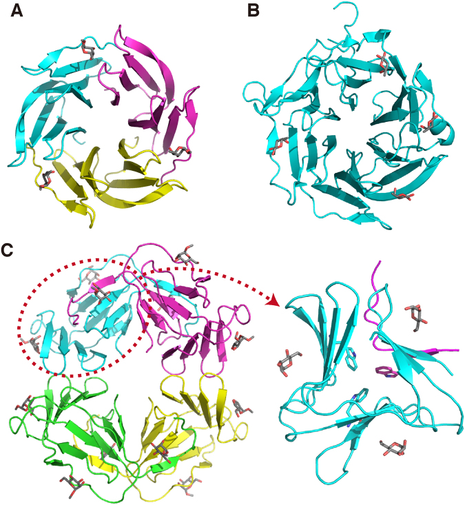
Comparison of related lectin structures. (A) The crystal structure of RSL trimer in complex with methylfucose (PDB ID: 2BT9)18. (B) The crystal structure of AAL, consisting of a single polypeptide, in complex with fucose (PDB ID: 1OFZ)19. (C) The crystal structure of GNA tetramer in complex with methylmannose (PDB ID: 1MSA)20. In the right panel, a monomeric part of GNA with a swapping of a β-strand (circled in the left panel) is focused. In these figures, different polypeptide chains are distinguished by colors, and bound sugars are shown in stick representations. In the right panel of (C), a triad of Trp residues in the core is indicated also by stick.

