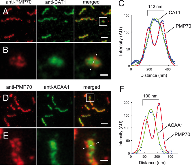
Figure 2.
Two-color STED nanoscopy of peroxisome membrane and matrix. Dual immunofluorescence on human skin fibroblasts. (A) Monoclonal anti-PMP70 labeled with KK114 secondary label in red, polyclonal rabbit anti-Catalase (anti-CAT1) labeled with Atto594-coupled secondary in green, and a merge of both (right). (B) Blow-up of box in (A). (C) Gaussian fit of the line scan marked in (B). (D) Anti-PMP70 labeled with KK114-coupled secondary antibody in red, polyclonal rabbit anti-acetyl-CoA acyltransferase1 (anti-ACAA1) in green, and merged images of both. (E) Inset magnification of (D). (F) Gaussian fit of the line scan marked in (E). Images were smoothed by 3 × 3 average filter and linearly scaled. Scale bars 500 nm (A,D), and 100 nm (B,E).

