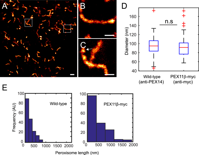
Figure 3.
STED sub-diffraction image of hyper-tubulated peroxisomes. Peroxisome proliferation was induced by overexpression of PEX11β. (A) STED overview image of HeLa cell overexpressing PEX11β-myc fusion 24 hours after transfection, probed with a monoclonal anti-myc antibody and labeled with a secondary antibody conjugated to KK114 dye. (B,C) Blow up of hyper-tubulated PEX11β-myc structures. Arrow indicates a vesicle that may be budding of from the peroxisome. (D) Size (diameter) distribution of wild-type and hyper-tubulated peroxisomes. PEX11β-myc tubule’s diameter (probed with anti-Myc) was measured by FWHM of the Gaussian fit (N = 200 line scans from 70 peroxisomes from five cells). The mean diameter (dmean) = 94.9 ± 21.8 nm for wild-type and (dmean) = 91.8 nm ± 20.6 nm (±SD) for PEX11β-myc. For comparison, untransfected HeLa cells probed with anti-PEX14, quantified by distance between peaks of Gaussian fit membrane profiles (N = 74 profiles from 16 independent slices). Differences in the peroxisomal mean diameter were not significant (n.s) with p > 0.05. Scale bars 500 nm. (E) Histogram of peroxisome length showing PEX11β-myc hyper-tubulation >800 nm (N = 169 peroxisomes). Control: HeLa cells probed with the anti-PEX14 show no (N = 142 peroxisomes).

