Fig. 6.
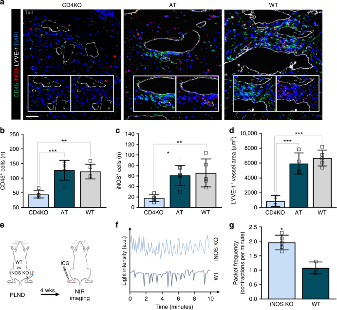
Adoptive transferred CD4+ T cells mediate iNOS production by macrophages after lymphatic injury. a Mice killed 6 weeks after tail skin and lymphatic excision and 1 week after the last adoptive transfer of CD4+ T cells for AT mice. Representative immunofluorescent images of tail cross-sections co-localizing LYVE-1+ initial lymphatic vessels with CD45 and iNOS; inset images demonstrate CD45 alone (inset left) and iNOS alone (inset right); scale bar, 50 µm. b, c Quantification of CD45+ (b) and iNOS+ cells (c) within 50 µm of LYVE-1+ lymphatic vessels (n = 5 per group; 4 hpf per mouse). d Quantification of LYVE-1+ lymphatic vessel area (n = 4 for CD4KO and AT groups, n = 5 for WT group; 4 hpf per mouse). e Schematic diagram of PLND in WT versus iNOS KO mice. Mice analyzed by NIR imaging 4 weeks after surgery. f Representative graphs of collecting lymphatic vessel pumping as determined by changes in ICG light intensity. g Quantification of collecting lymphatic vessel contractions per minute (n = 5 per group). Data representative of a minimum of two independent experiments with similar results; statistical analyses of one experiment shown. Mean ± s.d.; *P < 0.05, **P < 0.01, and ***P < 0.001 by one-way ANOVA with Tukey’s multiple comparisons test. AT, CD4KO mice that underwent adoptive transfer with naive CD4+ T cells; hpf, high-powered field; NIR, near-infrared; PLND, popliteal lymph node dissection

