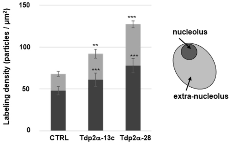
FIGURE 4.

Increased transcriptional activity is observed in the nucleolar and extra-nucleolar regions of MtTdp2α-overexpressing cells. Quantitative evaluation of anti-BrU immunolabeling. BrU (100 mM) was provided for 1 h to 4-day old M. truncatula cell suspension cultures (CTRL and MtTdp2α-overexpressing lines) and BrU incorporation into transcripts was measured in the nucleolar (dark gray) and extra-nucleolar (pale gray) regions. A schematic representation of the nucleus with highlighted nucleolar and extra-nucleolar areas is shown. np, nucleoplasm. The frequency of labeled transcripts is expressed as number of gold particles per μm2. Data represent the mean ± SD of three replicates per experiment. Asterisks indicate statistically significant differences determined using Student’s t-test (P < 0.05).
