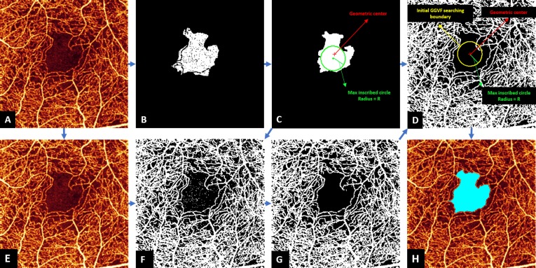
Figure 2.
Foveal avascular zone (FAZ) detection in the inner retina of an eye with NPDR eye. The original angiogram (A) is processed by seeded region growing from the center of the image to obtain the initial FAZ and ancillary nonperfusion areas (B), and morphologic operations removed regions of ancillary nonperfusion (C). Locating geometric center was applied to obtain a circle center coordinate (red dot in [C, D]), and extraction of maximum inscribed circle was applied to obtain a radius (green line segment in [C, D]), which was then used to generate the initial GGVF searching boundary (yellow circle in [D]). The original angiogram (A) is normalized to the range [0 1] (E) and processed by Otsu's N thresholding (F) and noise removal (G), which is used to create an edge map for GGVF model (G). The initial GGVF searching boundary was then applied on the edge map, and the GGVF active contour model was applied to identify the final FAZ. The resulting final FAZ (light blue) is overlaid on the original angiogram (H).

