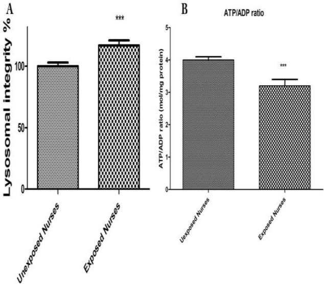
Figure 2.
Lysosomal membrane damage (A) and ATP level (B). Human lymphocytes (106 cells/mL) were suspended in the RPMI 1640 at 37 oC. Lysosomal membrane damage was determined as the difference in redistribution of acridine orange from lysosomes into cytosol between both groups. ATP/ADP ratio were determined by Luciferin /Luciferase assay System as described in Materials and Methods. Each bar represents mean SEM. Each group consisted of 50 individuals. *** p < 0.001 significant difference compared to the unexposed group

