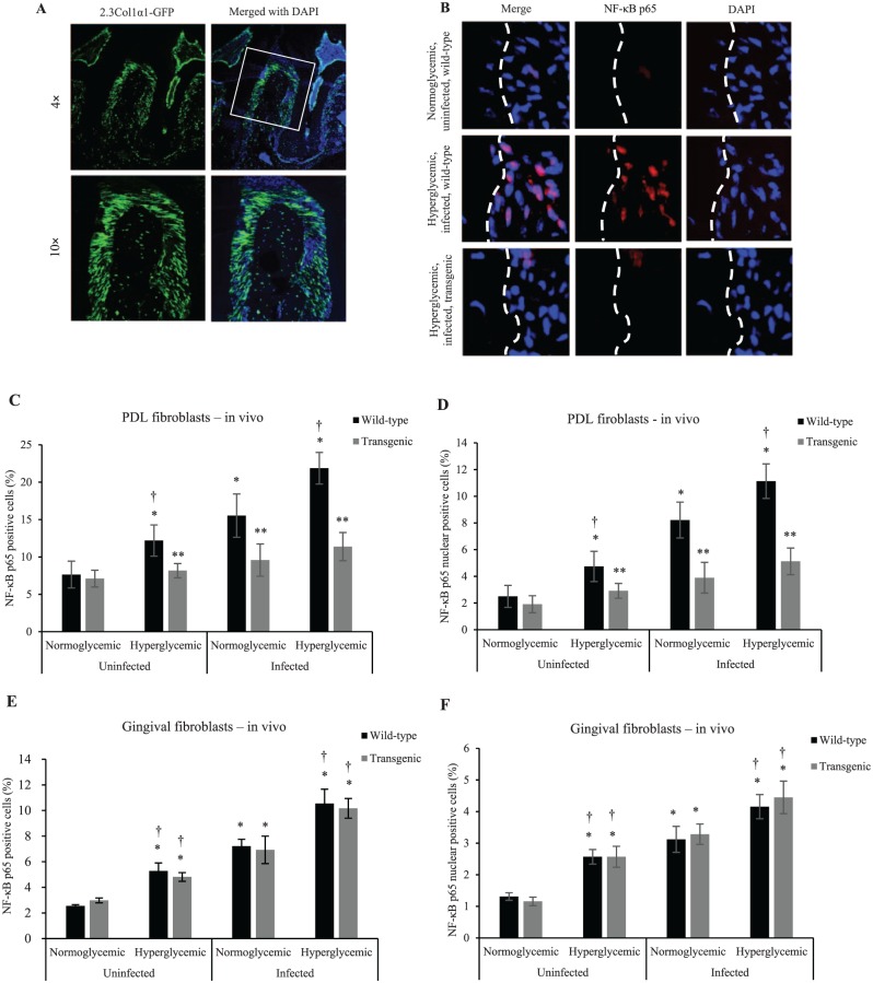
Figure 1.
Diabetes and oral infection induce NF-κB activation in PDL fibroblasts, which could be inhibited by the 2.3-kb Col1α1 promoter gene typical of osteoblast lineage cells. (A) Localization of GFP in periodontal tissue of 2.3Col1α1-GFP transgenic mice. 2.3Col1α1-GFP signal was detected in PDL and alveolar bone with expression by PDL fibroblasts, bone-lining cells, and osteocytes. (B) NF-κB p65 expression by PDL fibroblasts by immunofluorescence. (C) NF-κB p65 expression was measured in vivo by immunofluorescence as percentage of immunopositive fibroblastic cells in the PDL. (D) NF-κB p65 nuclear localization was measured in vivo by colocalization of NF-κB p65 (red) and DAPI (blue) nuclear staining and presented as the percentage of fibroblastic cells in the PDL with NF-κB p65 detected in the nucleus. (E) Total NF-κB p65 expression was measured by immunofluorescence in vivo and presented as the percentage of immunopositive fibroblastic cells in gingiva. (F) NF-κB p65 nuclear localization was measured in vivo by colocalization of NF-κB p65 (red) and DAPI (blue) nuclear staining and presented as the percentage of fibroblastic cells in the gingiva with NF-κB p65 detected in the nucleus. *P < 0.05 (vs. uninfected normoglycemic group). **P < 0.05 (vs. matched wild-type group). †P < 0.05 (vs. matched normoglycemic group). Col1α1, collagen type 1, alpha 1; GFP, green fluorescent protein; NF-κB, nuclear factor kappa B; PDL, periodontal ligament. Error bars indicate SEM.

