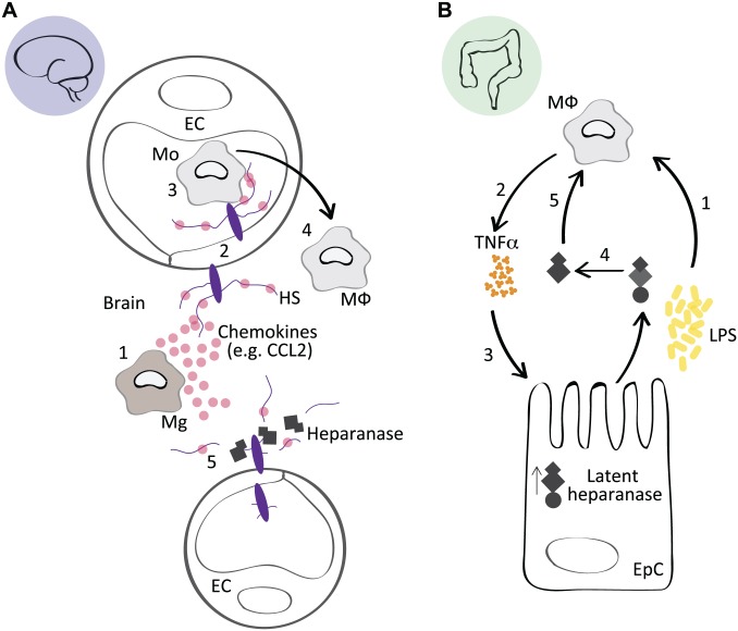
Figure 1.
Context-specific effects of heparanase during inflammation. (A) In response to a systemic administration of LPS, or a local inflammatory stimuli, for example, Aβ, activated Mg in the brain secrete chemokines such as CCL2 (1). HSPGs on EC bind CCL2 and transcytose the chemokine to the luminal surface of the endothelium (2), where it participates in the recruitment of circulating Mo (3) that transmigrate across the blood brain barrier, where they differentiate into MΦ and participate in the inflammatory response (4). In Hpa-tg mice, heparanase-mediated fragmentation of endothelial HPSGs suppressed the presentation of chemokines on the endothelial lumen, reducing the recruitment of monocytes, thus attenuating the inflammatory state in the brain. This model is adapted from Zhang et al.9 (B) The inflammatory condition ulcerative colitis is associated with impaired barrier function of epithelial cells in the colon (EpC), which leads to an increased exposure of MΦ to bacterial debris from the intestinal flora, including LPS (1). LPS induces TNFα and cathepsin-l expression in MΦ (2), which upregulates the latent form of heparanase in EpCs (3). Latent heparanase is activated through the action of macrophage-derived cathepsin-l (4). The active heparanase acts on the MΦ through an HS-dependent mechanism, potentially releasing TLR4 from an HS-mediated inhibition state (5), which prolongs the inflammatory activation of the MΦ and contributes to the persistence of colitis. This model is adapted from Lerner et al.35 Abbreviations: LPS, lipopolysaccharide; Aβ, amyloid-β; Mg, microglia; HSPGs, heparan sulfate proteoglycans; EC, endothelial cells; Mo, monocytes; MΦ, macrophages; EpC, epithelial cells in the colon; TNFα, tumor necrosis factor α; HS, heparan sulfate; TLR4, toll-like receptor 4.

