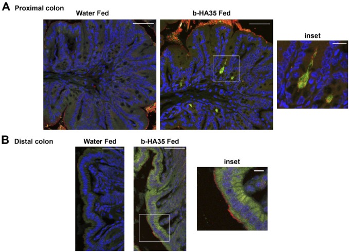
Figure 2.
HA35 induces β-defensin expression at colon epithelial cell surface. Co-localization of biotinylated HA35 (green) and β-defensin (red), co-localization (yellow) was detected in proximal colon (Panel A) or distal colon (Panel B) cross sections from mice fed water or 300 µg of biotinylated HA35 (b-HA35). Immunohistochemical staining with streptavidin 488 for HA35 and rabbit anti-murine β-defensin, goat anti-rabbit AlexaFluor 568 was performed as described in the “Materials and Methods” section. Nuclei were stained with VectaShield containing DAPI (blue). Image captured with Leica confocal and Image-Pro Plus software. Panel A scale bars are 50 µm and inset 5 µm. Panel B scale bars are 50 µm and inset 10 µm. Abbreviations: HA35, 35 kDa hyaluronan; DAPI, 4′,6-diamidino-2-phenylindole, dihydrochloride.

