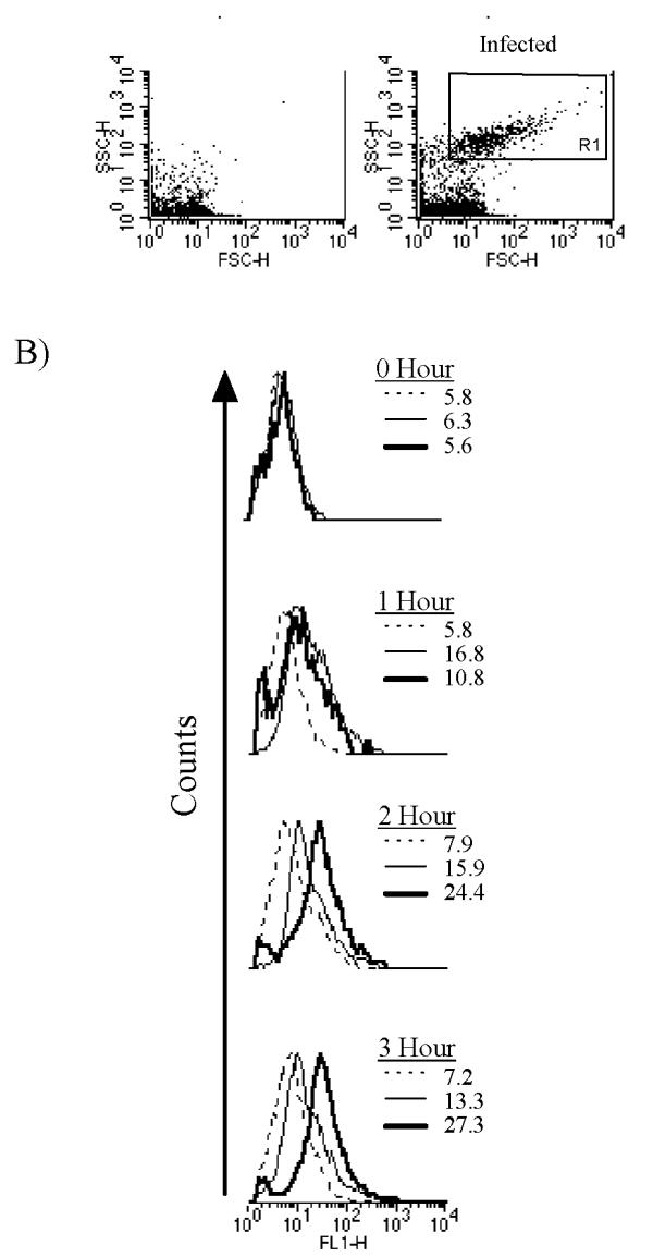
Figure 2.

GFP levels in the yopE::gfp strain either in the absence or presence of eukaryotic cells. (A) Forward (x-axis) and side (y-axis) light scattering plots of lysates made from either uninfected (left) or yopE::gfp-infected (right) RAW cells. (B) FL1 signals (green channel) of Rl-gated events (shown in [A]) either in yopE::gfp cultures diluted into tissue culture media (26°C-dotted line; 37°C-thin solid line) or of lysates made from RAW cells infected with yopE::gfp at a multiplicity of infection (MOI) of 20 (thick solid line). The mean fluorescence intensity of the size-gated bacteria from each culture is shown.
