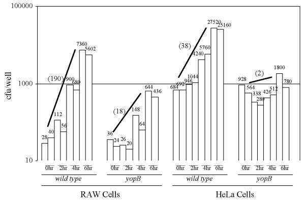
Figure 3.

Infection of cultured eukaryotic cells with the wild-type and yopB strains of Y. pseudotuberculosis. RAW macrophage-like cells (left) or HeLa cells (right) were infected with the indicated strain at a MOI of either 0.04 (left) or 0.4 (right). At the indicated times after the removal of unattached bacteria, the number of cell-associated bacteria per well was determined by viable plating. Shown both graphically and numerically are the number of colony-forming units (cfu) of two independent wells per time point. In parenthesis is the average fold-increase of each strain after the 6-hour infection period.
