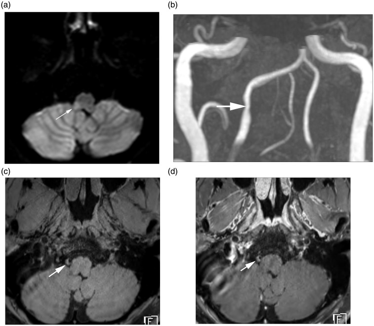
Figure 1.
HR-MRI findings of intracranial VAD with AIS. (a) Diffusion MRI demonstrates a focal acute infarction (arrow) involving right sided medulla. (b) TOF MR angiography reveals focal stenosis (arrow) at V4 segment of the right vertebral artery. (c) Axial pre-contrast T1-weighted MR image demonstrates crescentic T1 hyperintensity (arrow) at the stenotic segment, suggesting intramural hematoma. (d) Axial post-contrast T1-weighted image demonstrates perivascular enhancement (arrow) along the affected vessel.
HR-MRI: high-resolution magnetic resonance imaging; VAD: vertebral artery dissection; AIS: acute ischemic stroke; TOF: time of flight; MR: magnetic resonance.

