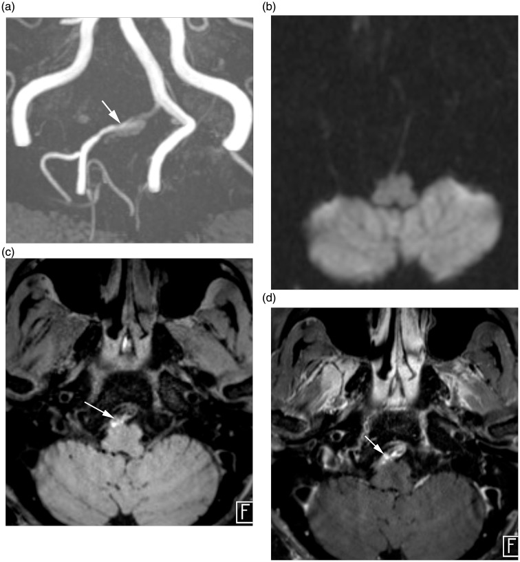
Figure 2.
HR-MRI findings of intracranial VAD without AIS. (a) TOF MR angiography demonstrate aneurysmal dilation (arrow) at V4 segment of the right vertebral artery. (b) Diffusion MRI reveals no evidence of diffusion restricted lesion involving brain parenchyma. (c) Axial pre-contrast T1-weighted image demonstrates intimal flap and hyperintense intramural hematoma (arrow). (d) Axial post-contrast T1-weighted image demonstrates perivascular enhancement (arrow) along the affected vessel.
HR-MRI: high-resolution magnetic resonance imaging; VAD: vertebral artery dissection; AIS: acute ischemic stroke; TOF: time of flight; MR: magnetic resonance.

