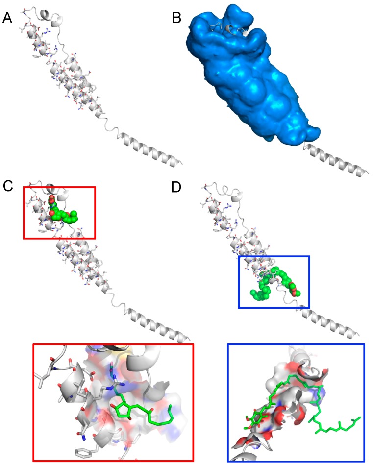
Figure 2.
Binding modes of unoprostone and ubidecarenone to the membrane-fusion subunit from the Ebola virus envelope glycoprotein, GP2 (PDB identifier 1ebo, chain F). (A) Binding site residues (stick representation) predicted by COFACTOR by comparing binding motifs to a library of the PDB ligand-bound structures; (B) Search space for CANDOCK to dock fragments of small molecule compounds that are reconstructed while incorporating flexibility of both small molecule and the protein; (C) Docked conformation of uniprostone bound to the region of the fusion peptides forming disulfide-bonded loop that is homologous to an immunosuppressive sequence in retroviral glycoproteins. Uniprostone is shown as spheres (top) along with its interaction to the protein surface up to 10 Å (bottom); (D) Docked conformation of ubidecarenone bound to coiled coil region near the C-terminal end that acts as the membrane anchor. Ubidecarenone is shown as spheres (top) along with its interaction to the protein surface of the coiled coil region up to 10 Å (bottom). Together, these two molecules likely disrupt the conserved disulfide-bonded loop and the linker region that function as a hinge, transferring information from the GP1 receptor binding to trigger a conformational change in GP2, thereby disrupting the membrane fusion event.

