
Figure 4.
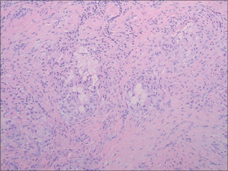
Within the dermis, there are somewhat nodular collections of lipid-laden histiocytes highlighted by a CD68 stain. Areas of extracellular lipid deposition are also seen including cholesterol clefts. (H and E, ×100)
Official websites use .gov
A
.gov website belongs to an official
government organization in the United States.
Secure .gov websites use HTTPS
A lock (
) or https:// means you've safely
connected to the .gov website. Share sensitive
information only on official, secure websites.
Within the dermis, there are somewhat nodular collections of lipid-laden histiocytes highlighted by a CD68 stain. Areas of extracellular lipid deposition are also seen including cholesterol clefts. (H and E, ×100)