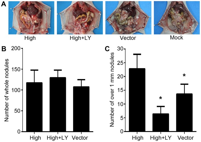
Figure 4.
PRL-3 promotes peritoneal spreading via the PI3K/AKT signaling pathway in vivo. (A) Representative peritoneal dissemination of SGC7901 cells in LY294002-treated mice. Mice were sacrificed 4 weeks post-injection and the peritoneum was observed. (B) The total number of metastatic nodules on the peritoneal surface in each treatment group. (C) The number of metastatic nodules >1 mm3 in each treatment group. The number of metastatic nodules was significantly lower in the high+LY group. The bars represent the mean ± standard deviation (n=6/group). *P<0.05 vs. the control group. Mock, normal nude mice; vector, SGC7901 transfected with control empty vector; high, SGC7901/EGFP-PRL-3 cells; high+LY, plasmid SGC7901/EGFP-PRL-3/LY294002 cells. PRL-3, phosphatase of regenerating liver-3; AKT, RAC serine/threonine-protein kinase; PI3K, phosphatidylinositol 3-kinase.

