
Figure 1.
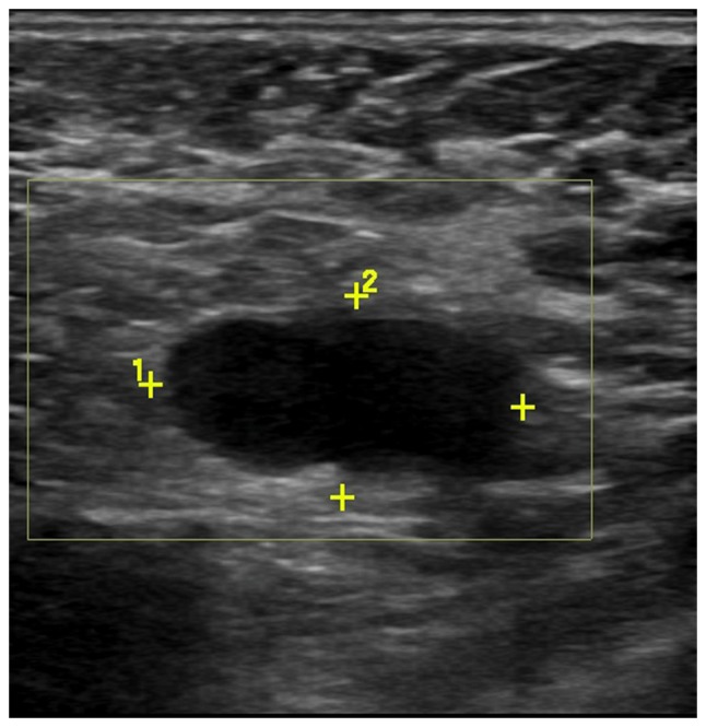
Scans of the left axillary lymph node from a 46-year-old female patient with invasive ductal carcinoma. Grayscale sonography showed diffusely enlarged lymph node with effaced fatty hilum.
Official websites use .gov
A
.gov website belongs to an official
government organization in the United States.
Secure .gov websites use HTTPS
A lock (
) or https:// means you've safely
connected to the .gov website. Share sensitive
information only on official, secure websites.
Scans of the left axillary lymph node from a 46-year-old female patient with invasive ductal carcinoma. Grayscale sonography showed diffusely enlarged lymph node with effaced fatty hilum.