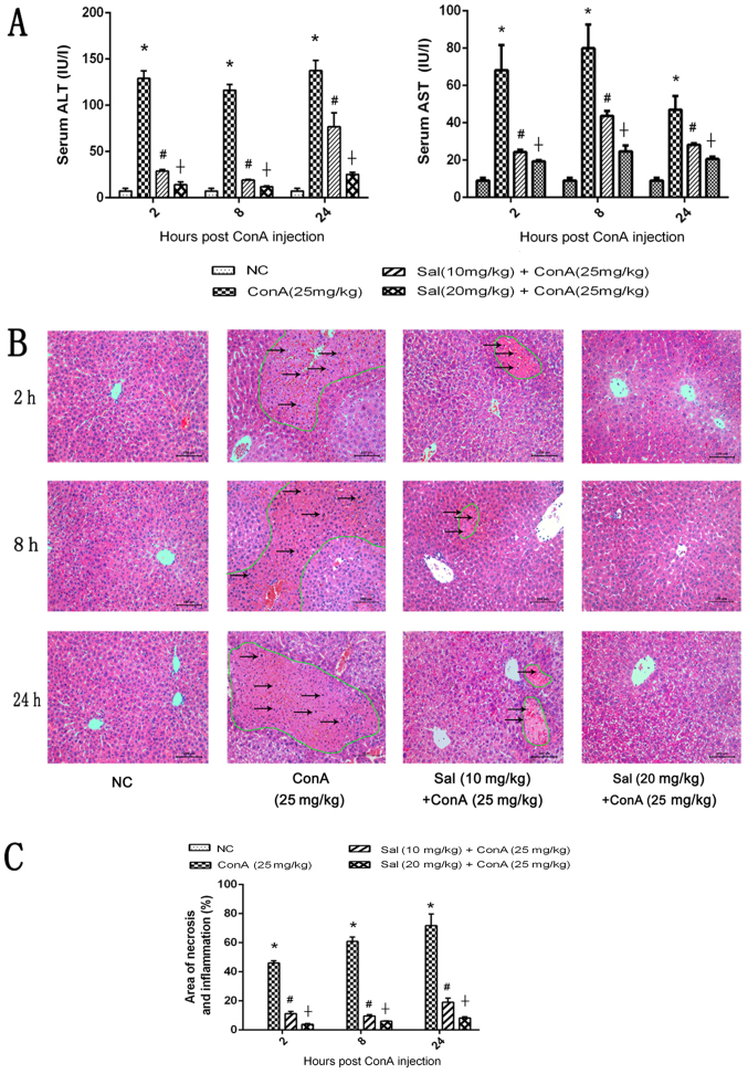
Figure 2.
Effects of Sal on liver function and pathology of mice in ConA-induced liver injury. (A) Levels of serum ALT and AST. (B) H&E staining of liver sections (original magnification, ×200). There were extensive areas of necrosis, which were eosinophilic and disorganized (circled by green lines), and an increased inflammatory response (indicated by black arrows) in liver tissues treated with ConA alone, and these effects increased over time. However, Sal treatment attenuated these effects at the three time points, indicated by the decreased areas of necrosis. (C) Necrotic and edema areas in H&E-stained sections were analyzed with Image-Pro Plus software 6.0. Data are presented as the mean + standard deviation (n=6). *P<0.05 vs. NC; #P<0.05 vs. ConA (25 mg/kg); †P<0.05 vs. Sal (10 mg/kg) + ConA (25 mg/kg). Sal, salidroside; ConA, concanavalin A; NC, negative control; H&E, hematoxylin and eosin; ALT, alanine aminotransferase; AST, aspartate aminotransferase.

