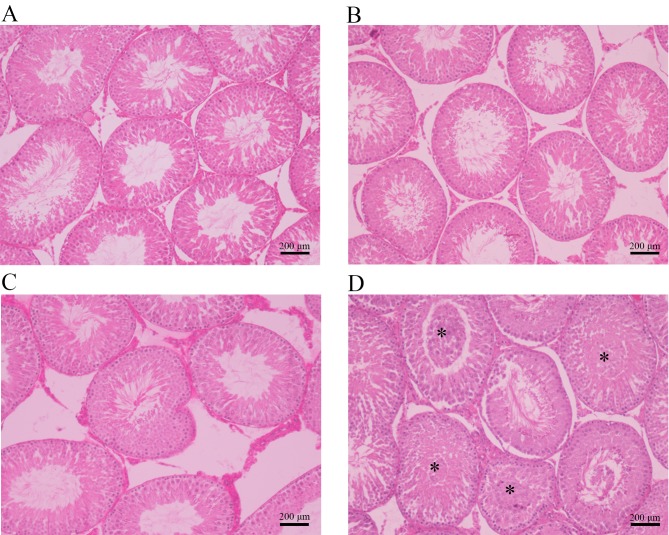
Figure 3.
Histopathological photomicrographs of cross-sections of testis samples from the (A) control, (B) 20 mg/kg cimetidine, (C) 40 mg/kg cimetidine and (D) 120 mg/kg cimetidine groups (hematoxylin and eosin staining; magnification, ×100; scale bar, 200 µm). The control, 20 mg/kg cimetidine and 40 mg/kg cimetidine groups displayed a compact and regular arrangement of cells in the seminiferous tubules. The 120 mg/kg group exhibited cell material shedding in the lumen of seminiferous tubules (asterisks).

