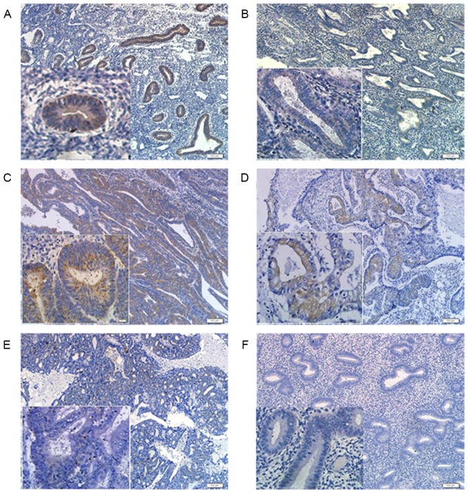
Figure 3.
Representative images of immunohistochemical analysis of PDCD4 in the EEC and control groups (×100 magnification and ×400 magnification; scale bars=200 µm). (A) Immunohistochemical staining of PDCD4 in the proliferative phase of control endometrium. (B) Immunohistochemical staining of PDCD4 in the secretory phase of control endometrium. (C) Immunohistochemical staining of PDCD4 in G1 EEC tissues. (D) Immunohistochemical staining of PDCD4 in G2 EEC tissues. (E) Immunohistochemical staining of PDCD4 in G3 EEC tissues. (F) Negative control. PDCD4, programmed cell death 4; EEC, endometrioid endometrial carcinomas.

