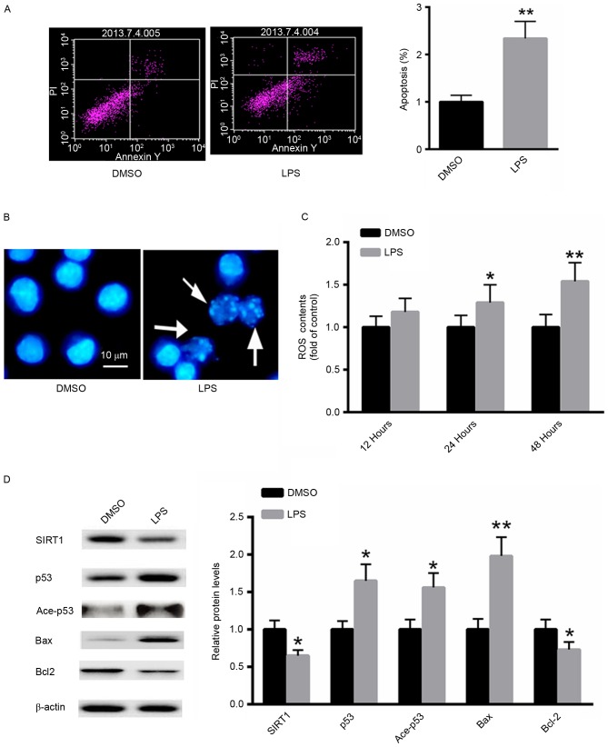
Figure 2.
LPS induces apoptosis in A549 cells. (A) Flow cytometric analysis. (B) Hoechst 33258 staining. (C) Detection of ROS generation. (D) Western blot analysis. *P<0.05, **P<0.01 vs. the untreated control. LPS, Pseudomonas aeruginosa lipopolysaccharide; DMSO, dimethyl sulfoxide; PI, propidium iodide; Ace, acetylated; ROS, reactive oxygen species; SIRT1, sirtuin 1; Bcl-2, B-cell lymphoma 2; Bax, Bcl-2-associated X protein.

