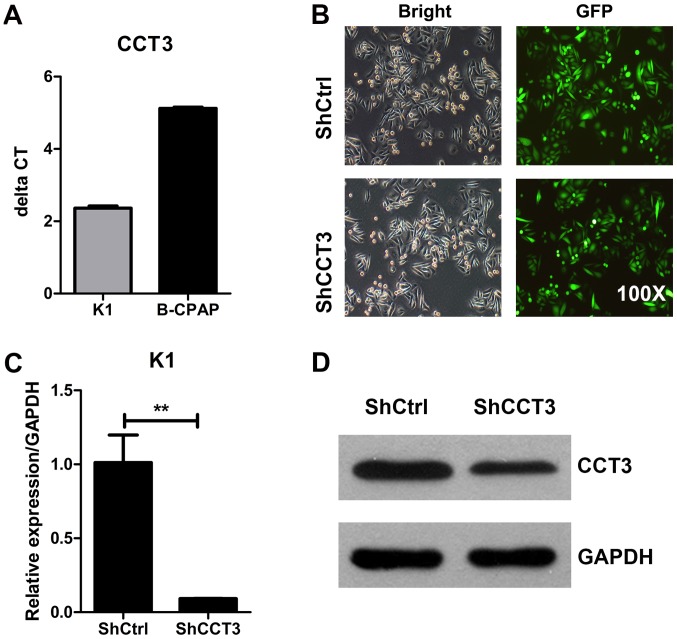
Figure 2.
Knockdown of CCT3 in human papillary thyroid carcinoma cells. (A) The 2−ΔΔCq value indicating the relative expression of CCT3 in K1 and B-CPAP cells. (B) Infection efficiency as determined by light and fluorescence microscopy at 72 h Following lentiviral infection in K1 cells. Original magnification, ×200. Representative images of the cultures are shown. (C) At 5 days post-infection, CCT3 mRNA levels in K1 cells were measured by reverse transcription-quantitative polymerase chain reaction and normalized to GAPDH. n=3; data points are presented as the mean ± standard error of the mean. **P<0.01, shCtrl vs. shCCT3. (D) CCT3 protein expression was analyzed by western blot analysis in the shCtrl and shCCT3 K1 cells. GAPDH was used as an internal control. CCT3, chaperonin containing TCP1 subunit 3; shCtrl, control short hairpin RNA.

