Figure 3.
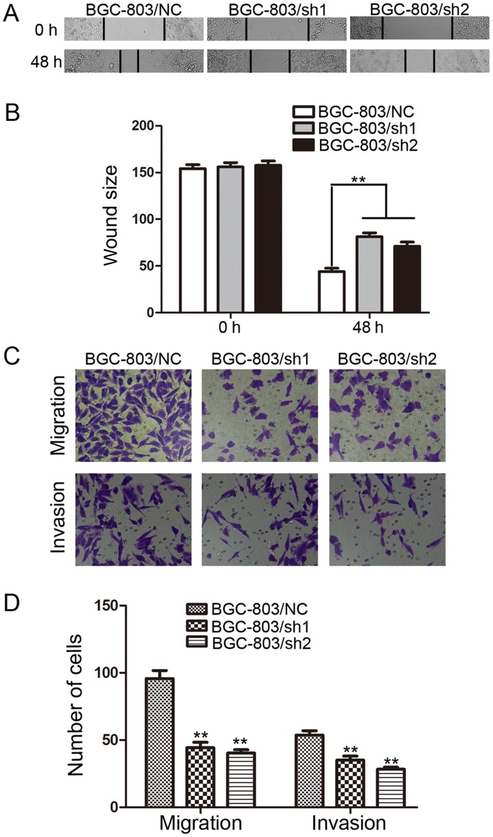
EHMT1 promotes the migration and invasion of GC cells. (A) BGC-803 cells were transfected with EHMT1 shRNA1/2 or NC shRNA, and representative images of wound healing assays were captured at 0 and 48 h post-wounding (magnification, ×200). (B) Quantification of the distance between wound edges of BGC-803/NC, BGC-803/sh1 and BGC-803/sh2 cells. (C) Representative images of migratory and invasive cells that traversed the micropore membrane following transfection with crystal violet staining (magnification, ×200). (D) Average number of migratory cells and invasive cells from 5 fields of view. Data are represented as the mean ± standard deviation of 3 independent experiments. **P<0.01 vs. control. EHMT1, euchromatic histone lysine methyltransferase 1; GC, gastric cancer; shRNA, short hairpin RNA; NC, negative control; sh1, EHMT1 shRNA 1; sh2, EHMT1 shRNA 2.

