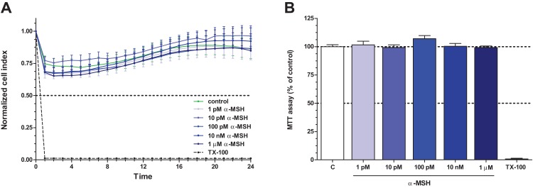
Figure 2. The effect of different concentrations of α-MSH on the cell viability of rat brain endothelial cells.
(A) Cultured brain endothelial cells were treated with α-MSH (1 pM to 1 μM) for 24 h. Control group received culture medium. Endothelial cells treated with 1% Triton X-100 detergent were used as cytotoxicity control. The cell index followed by impedance did not change after α-MSH treatment. (B) The metabolic activity of rat brain endothelial cells was measured by MTT assay, which did not detect any alteration caused by α-MSH. Mean ± SEM, n = 4–8. C, control group; TX-100, Triton X-100.

