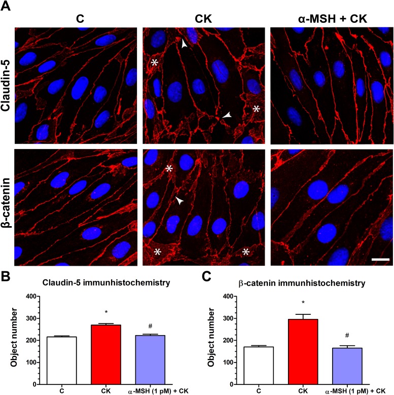
Figure 5. The effect of α-MSH treatment on the immunostaining of tight junction proteins claudin-5 and β-catenin in cytokine treated rat brain endothelial cells.
(A) Rat brain endothelial cells were treated with cytokines (10 ng/ml TNF-α and 10 ng/ml IL-1β) without or with 1 pM α-MSH for 1 h and stained for tight junction proteins claudin-5 and β-catenin. The control group received culture medium. The fluorescent microscopy images demonstrate the expression and organization of the junctional proteins. Cytokine treatment significantly increased the number of gaps and intracellular redistribution compared to the control and α-MSH treated group. Arrowheads: gaps and fragmented junctional staining; asterisks: cytoplasmic redistribution of junctional proteins. Scale bar: 10 μm. (B) The object number on the claudin-5 immunostained pictures was quantified by MATLAB software. (C) The object number on the β-catenin immunostained pictures was quantified by MATLAB software. Mean ± SEM, n = 4, *P < 0.05, #P < 0.05. *: CK compared to C; #: CK + MSH compared to CK. C, control group; CK, cytokine treated group; α-MSH + CK, α-MSH, and cytokine treated group.

