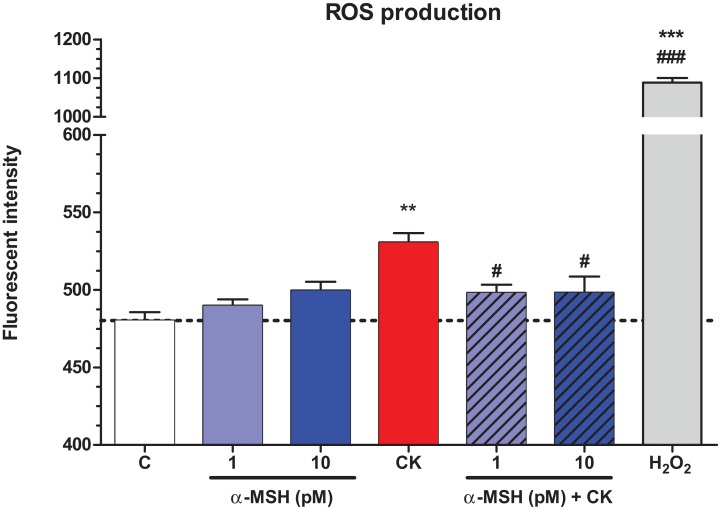
Figure 6. The effect of α-MSH treatment on the reactive oxygen species production in cytokine treated rat brain endothelial cells.
Rat brain endothelial cells were treated with cytokines (10 ng/ml TNF-α and 10 ng/ml IL-1β) without or with 1 or 10 pM α-MSH for 1 h. Cells were also treated with 1 or 10 pM α-MSH alone. Control group received culture medium. Mean ± SEM, n = 4–8. C, control group; CK, cytokine treated group; H2O2, hydrogen peroxide treated group (100 μM). **P < 0.01, ***P < 0.001, #P < 0.05, ###P < 0.001. Asterisks indicate that groups were compared to the control group. Pound signs indicate that groups were compared to the cytokine-treated group. C, control group; CK, cytokine treated group; α-MSH+CK, α-MSH and cytokine treated group.

