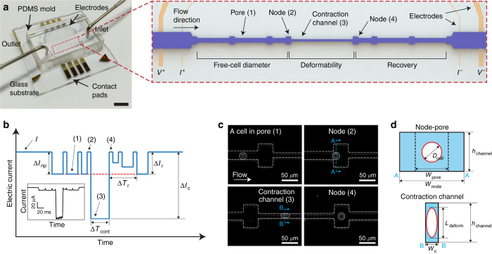
Figure 1.
Principle of mechanical phenotyping via mechano-NPS. (a) A photographic image of the microfluidic platform. The scale bar corresponds to 4 mm. Red-dashed box shows a close-up view of the entire microfluidic channel. The microfluidic channel (pore) is segmented by nodes and a contraction channel. Two electrodes at both ends of the channel apply a constant voltage (1 V), and two inner electrodes measure the change of current across the channel. The regions where free-cell diameter, deformed diameter, and cell recovery are measured are as indicated. (b) Expected current pulse generated by a cell transiting the microfluidic channel. I, ΔInp, ΔIc, and ΔIr correspond to the baseline current and the current drop by a cell transiting a node-pore, a contraction channel, and a node-pore after the contraction channel, respectively. Numbers in parentheses (1–4) correspond to the same specific segments of the microchannel (pore, node, and contraction channel) in (a). ΔTcont corresponds to the time duration of a cell passing through the contraction channel, and ΔTr indicates the time needed for ΔIr to equal ΔInp (see Supplementary Figure S6 for detailed information). (inset) An actual current pulse caused by a human mammary epithelial cell traversing the channel. (c) Time-snapshots of an MCF-7 cell (bordered by a white circle) in each of the different segments of the microfluidic channel (white dashed line; see Supplementary Videos 1 and 2 for detailed information). Numbers in parentheses (1–4) correspond to the same specific segments of the microchannel (pore, node, and contraction channel) in (a). (d) Cross-sectional diagram of the channel segments occupied by a cell. ‘AA’ and ‘BB’ indicate the corresponding cross-sections in (c). wpore, wnode, wc, and hchannel correspond to the widths of the pore, node, and the contraction channel, and the height of the channel, respectively. Dcell and Ldeform correspond to the free-cell diameter in the node-pore channel and the elongated length of the deformed cell in the contraction channel, respectively.

