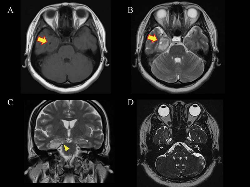
Figure 2.

Magnetic resonance imaging (MRI) studies. Axial images show (A) isointensity (arrow) in T1-weighted and (B) hyperintensity (arrow) in T2-weighted images. (C) A coronal T2-weighted image shows extension of the tumour to the cerebral peduncle (arrowhead). (D) Constructive interference in steady-state MRI demonstrates no tumour involvement on the facial nucleus or facial nerve.
