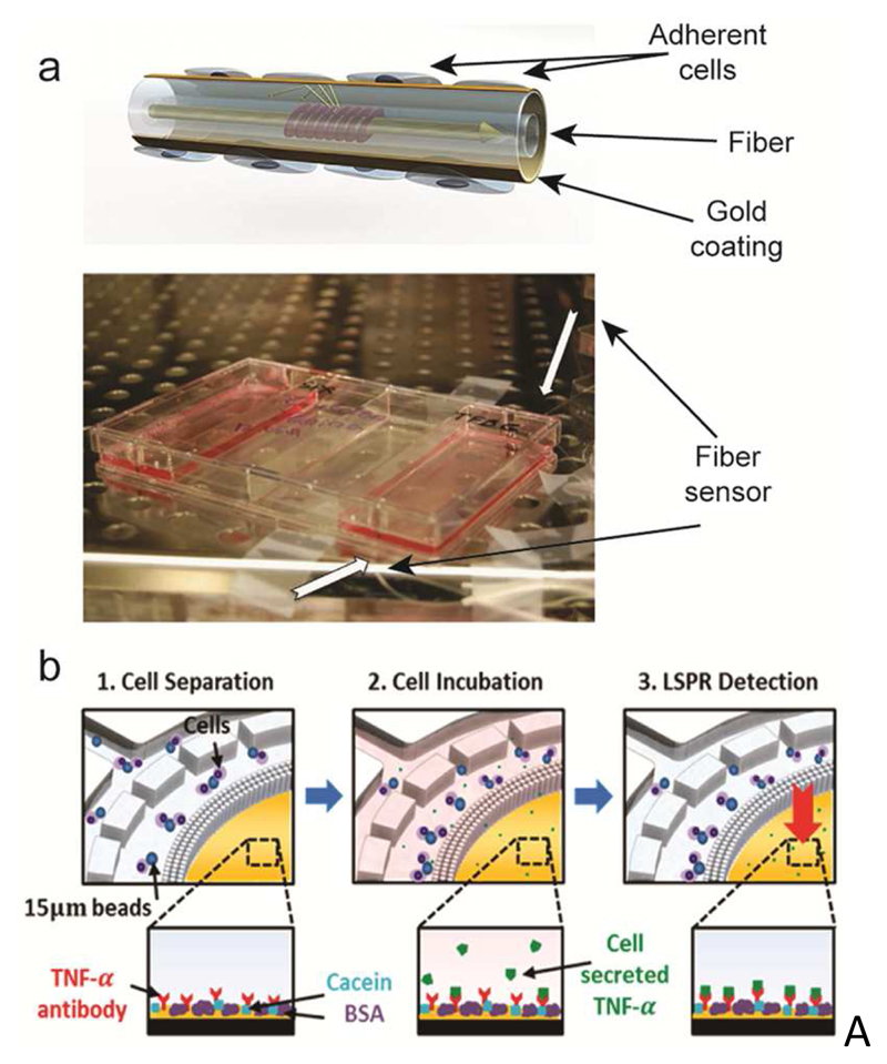
Figure 5.
SPR-based sensors. a) A SPR fiber sensor for real-time monitoring of cellular responses, cultured in standard cell-culture flasks. The schematic shows the working principle of the sensor. Cells are cultured on the gold-patterned fiber surface. Light traveling through the waveguide interacts with the adherent cells on the fiber. The photograph shows the integration of the sensor in a cell-culture flask. Adapted with permission from Shevchenko et al., Biosens. Bioelectron. 56, 359-367.174 Copyright 2014. b) LSPR-based detection of cytokine (TNF-α) secretion by immune cells isolated from human blood. Functionalized beads bind to the target cells. (1) The sample then flows through a microfluidic chamber featuring a sieve structure to selectively retain the cells that are bound to beads. (2) Cells are then stimulated to induce cytokine secretion. (3) Cytokine secretion is quantified by the on-chip LSPR sensor functionalized with target-specific antibodies. The insets at the bottom show the LSPR-sensor surface at different analysis stages. Reprinted with permission from Oh et al., ACS Nano 8, 2667-2676.181 Copyright 2014.

