
Figure 1. Regional brain vulnerability to hypoglycemia is predicted by baseline glucose demand.
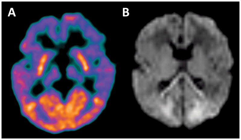
(A) FDG-PET brain imaging was performed in a 7 month old for the purposes of epilepsy source localization. This shows the well documented finding of elevated FDG uptake in the occipital lobes during early infancy. (B) Brain MRI including diffusion weighted imaging (DWI) was performed on an infant a few days after experiencing severe refractory hypoglycemia. This DWI image shows areas of diffusion restriction, representing focal brain injury, predominantly in the medial occipital lobes. An additional area of diffusion restriction in the splenium of the corpus callosum was interpreted to represent developing secondary Wallerian degeneration arising from the occipital lobe injury.