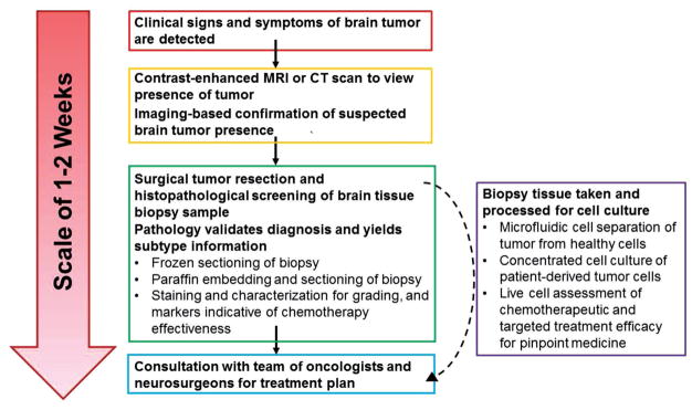
Figure 2.
Timescale of brain tumor detection and diagnosis methods including where microfluidic diagnostic methods could improve time to diagnosis and accuracy in prognosis. Conventionally, the process of a brain tumor diagnosis begins with presentation of symptoms and imaging-based assessment of lesion presence. Upon imaging-based confirmation of brain tumor presence, an invasive surgical procedure is scheduled to procure tissue biopsies for histopathological analyses. The pathology report containing information about the formal cancer diagnosis and staging can often take several weeks to generate, even when expedited. Microfluidic platforms provide an opportunity for the real-time analysis of tissue samples and the rapid processing of liquid biopsies (such as blood or serum samples), bypassing the potentially lengthy waiting time for diagnosis, staging information, or mutation status of GBM. MRI: Magnetic resonance imaging; CT: Computed tomography scan.

