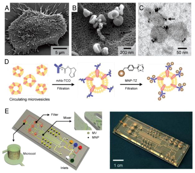
Figure 5.
Human glioblastoma cells produce abundant microvesicles (MVs), which can be analyzed by micronuclear magnetic resonance (μNMR). A) Scanning electron microscopic image of a primary human glioblastoma cell (GBM20/3) grown in culture, releasing abundant MVs. B) High-magnification image shows that many of the MVs on the cell surface assumed typical saucer-shaped characteristics of exosomes. C) Transmission electron microscopic image of MVs (≈80 nm) targeted with magnetic nanoparticles (MNPs) via CD63 antibody. The samples were purified by membrane filtration to collect small MVs. The MNPs appear as black dots (indicated by an arrow). D) Labeling procedure for extravesicular markers. The two-step BOND-2 assay configuration uses bioorthogonal amplification chemistry to maximize MNP binding onto target proteins on MVs (not drawn to scale). E) Microfluidic system for on-chip detection of circulating MVs. The system was designed to (i) allow MNP targeting of MVs, (ii) concentrate MNP-tagged MVs while removing unbound MNPs, and (iii) provide in-line μNMR detection. Reproduced with permission.[75] Copyright 2012, Nature Publishing Group.

