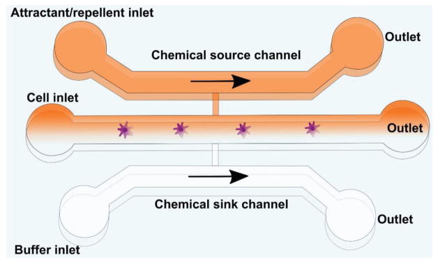
Figure 7.
General template of a microfluidic platform for chemotaxis studies. In the body, cells are exposed to extracellular chemoattractant gradients in 3D space and time, which influence cell behavior and migration. Simple microfluidic channels as depicted here can be used to present defined gradients of chemokines to cell types of interest, using either diffusion or laminar flow mixing to introduce chemokines into the outlets through a source channel in order to study glioma cell response to chemoattractants.

