Figure 1.
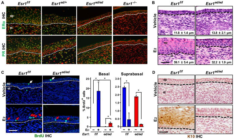
Epithelial ERα-deficient cervical epithelium partially responds to E2. (A) Specific ablation of ERα expression in the cervical epithelia. The female reproductive tracts were harvested from Esr1f/f and Esr1ed/ed mice at 8–10 weeks of age. Cervical sections were stained for ERα (green, upper panel) and PR (green, lower panel). Nuclei were stained with Hoechst 33258 (pseudocolored red). Esr1 null cervix (Esr1−/−) was used as negative control. Dotted lines separate epithelium (ep) from stroma (st). (B) Ovariectomized Esr1ed/ed mice were treated with vehicle or E2 for 7 days. Shown are representative images of H&E-stained cervical tissues. The thickness of epithelium is shown as mean ± SEM (n = 3–5). (C) Left panel: cervical sections described in (B) were stained for BrdU (green). Nuclei were stained with Hoechst 33258 (blue). BrdU+ basal and suprabasal cells are indicated by red and white arrows, respectively. Right panel: More than 200 cells per basal and suprabasal layer were counted. Results are shown as mean ± SEM (n = 3). *P < 0.05 (one-sided Wilcoxon rank sum test). (D) Staining for K10 (brown). Nuclei were counterstained with hematoxylin. Scale bar = 50 μm for (A), (C) and (D); 30 μm for (B)

