Figure 2.
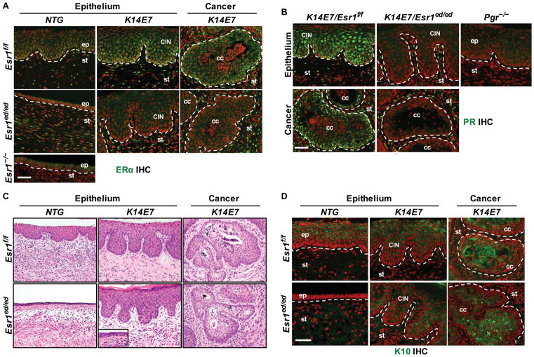
E2 promotes ERα − CxCa in K14E7/Esr1ed/ed mice. (A) Cervical sections of CxCa arising in K14E7/Esr1ed/ed mice stained for ERα (green). An Esr1−/− tissue section was used as negative control. Dotted lines separate stroma (st) from normal epithelium (ep), dysplastic epithelium (CIN), and cancer epithelium (cc). (B) PR staining (green). A Pgr−/− tissue section was used as negative control. (C) Representative H&E staining. Note that the nondiseased epithelia in K14E7/Esr1ed/ed mice were hypoplastic (inset). (D) K10 staining (green). In (A), (B) and (D), nuclei were stained with Hoechst 33258 (pseoudocolored red). Scale bar = 50 μm.

