
Fig. 2.
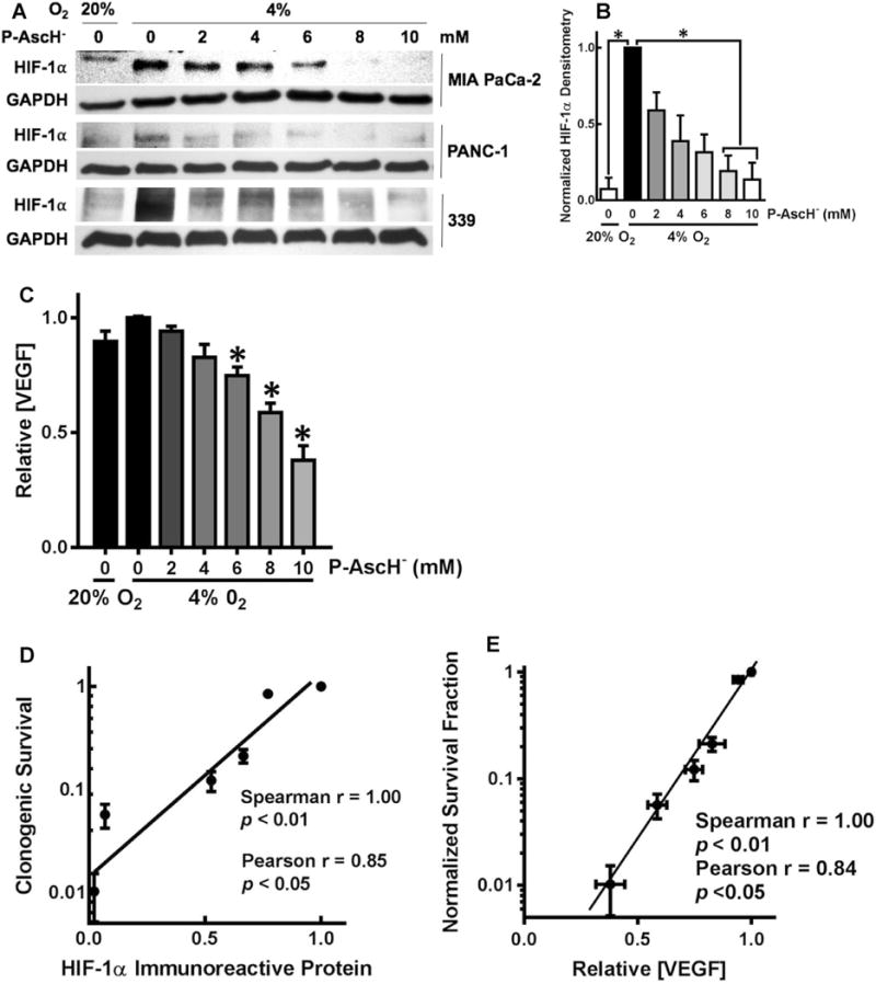
P-AscH− suppresses HIF-1α immunoreactive protein and VEGF secretion. a MIA PaCa-2, PANC-1, and 339 cell lines showed a dose-dependent decrease in HIF-1α expression after 1-h exposure to P-AscH−. Both cell lines had induction of HIF-1α immunoreactive protein in hypoxia. b In the MIA PaCa-2 cell line, densitometric analysis of HIF-1α demonstrates a decrease in HIF-1α expression with increasing doses of P-AscH− (n = 3, p < 0.05). c Secreted VEGF in media after 1-h P-AscH− and incubation in 4% O2 also demonstrated a dose-dependent decrease (means ± SEM, n = 6, *p < 0.05). d Clonogenic survival correlated with HIF-1α immunoreactive protein in MIA PaCa-2 cancer cells treated with P-AscH− (2–10 mM). e Clonogenic survival correlated with VEGF secretion in MIA PaCa-2 cancer cells with P-AscH− (2–10 mM)