
Fig. 6.
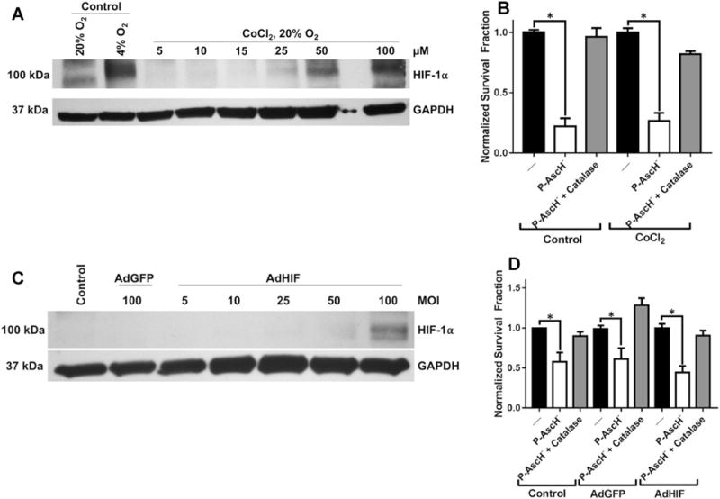
HIF-1α overexpression confers no resistance to P-AscH−. a Western blot of protein collected from MIA PaCa-2 cells incubated in normoxia with increasing concentration of CoCl2 (5–100 μM). Significant increases in HIF-1α protein are demonstrated at 50–100 μM CoCl2. b Clonogenic survival of MIA PaCa-2 cells pre-incubated for 8 h with CoCl2 (50 μM) prior to treatment with P-AscH− for 1 h demonstrates significant cytotoxicity (n = 3, p < 0.01) relative to non-P-AscH− controls. There were no significant differences in survival between P-AscH−-treated cells incubated with CoCl2 compared to untreated cells. c Western blot of protein collected from MIA PaCa-2 cells transfected with increasing MOI of AdHIF adenovirus. Significant overexpression of HIF-1α was demonstrated at 100 MOI. d Clonogenic survival of MIA PaCa-2 cells transfected with AdHIF adenovirus (100 MOI) treated with P-AscH− for 1 h in hypoxia. P-AscH− induced a significant cytotoxicity to AdHIF transfected cells (n = 3, p < 0.01) that was not significantly different from AdGFP and non-transfected controls