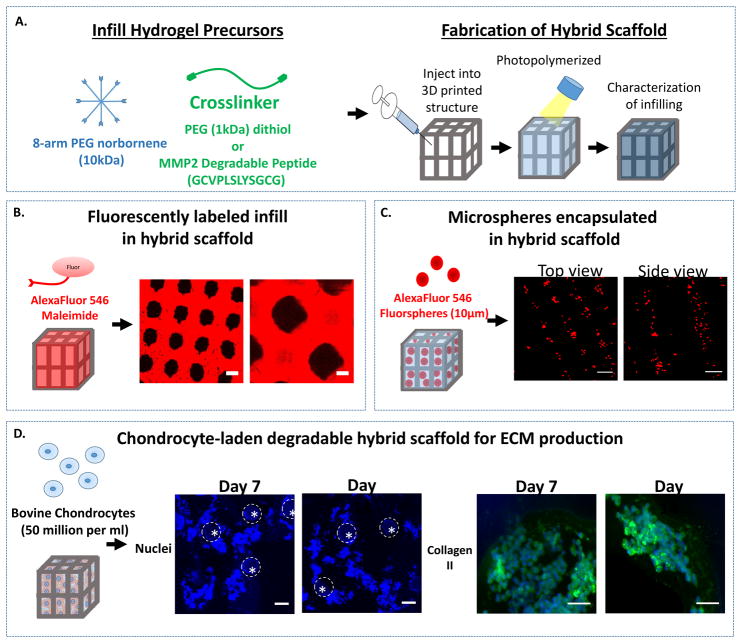
Figure 3.
A. A schematic of the photopolymerizable PEG precursor solutions for the infill and the fabrication of the hybrid scaffold by injecting the precursors and photopolymerizing. B. A schematic of the infilling of the hybrid scaffold with a fluorescently-labeled, PEG hydrogel. Representative confocal microscopy images shows successful infilling of the hydrogel (red) around the 3D printed support structure (black) (scale bar = 100 μm). C. A schematic of the infilling of the hybrid scaffold with fluorescently-labeled microspheres that are suspended in the infill solution and then subsequently photopolymerized to encapsulate them in the PEG hydrogel in the hybrid scaffold. Representative confocal microscopy images show the distribution of microspheres (red) through the top of the lattice (left) and a side view through the pillars (right) (scale bar= 100 μm). D. A schematic of the infilling of a chondrocyte-laden hybrid scaffold to which bovine chondrocytes were suspended in the infill solution and then subsequently photopolymerized to encapsulate them in the PEG hydrogel. Here a MMP2-sensitive PEG hydrogel was used. Cell nuclei (blue) shows chondrocytes are successfully infilled around the pillars (indicated by dotted line) of the hybrid scaffold at 7 and 14 days (scale bar = 100 μm). High magnification images of regions of high cell density where extracellular matrix neotissue is forming as shown by staining for collagen type II (green) and cell nuclei (blue) at 7 and 14 days (scale bar = 20 μm).

