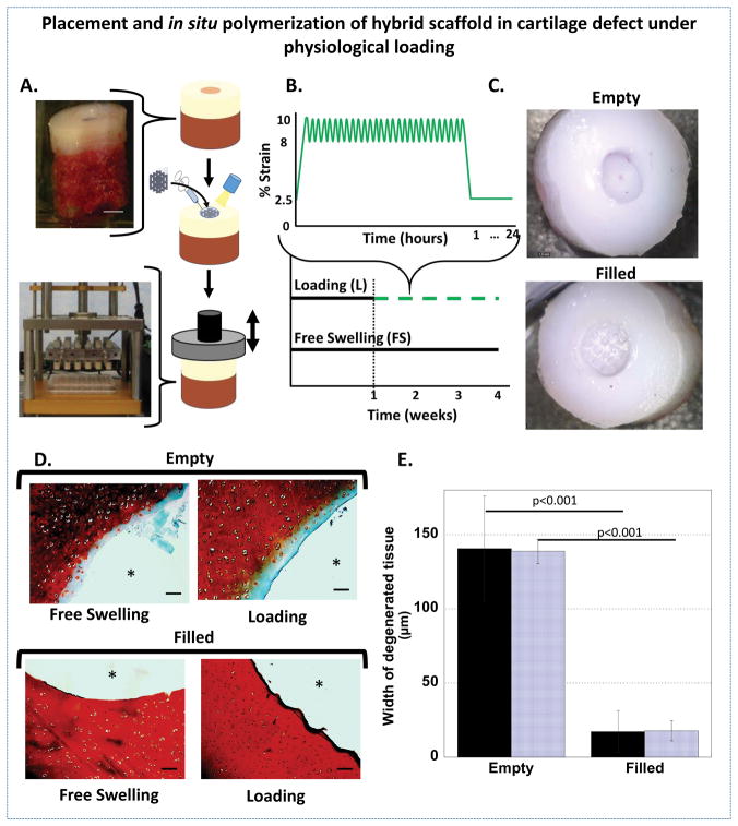
Figure 4.
A. A schematic showing the placement and infilling of the hybrid scaffold in a focal chondral defect of a porcine osteochondral plug (top) followed by intermittent, unconfined dynamic compression in custom bioreactors (bottom). B. A schematic of the intermittent dynamic loading profile (green, dashed) and free swelling (black, solid). Loading was applied by applying a slow ramp from the tare strain of 2.5% to 10% compressive strain followed by dynamic loading applied in a sinusoidal waveform at a frequency of 1 Hz between 8 and 10% compressive strain for one hour. A slow ramp was applied to remove the strain to 2.5% for 23 hours. C. Top view of osteochondral plugs with chondral defects that were left empty (top) and filled with the hybrid scaffold (bottom). Photographs were taken immediately after filling and prior to culture. D. Representative histological images of safranin O/fast green stained sections after 4 weeks show depletion of sGAGs (red) adjacent to the defect site in empty defects (* indicates empty defect site) and retention of sGAGs in the filled defects (* indicates hybrid scaffold) (scale bar = 100μm). E. The width of the degenerated tissue under free swelling (solid) and loading (striped) from semi-quantitative analysis of the sGAG histology (n=5, error bars = standard deviation).

