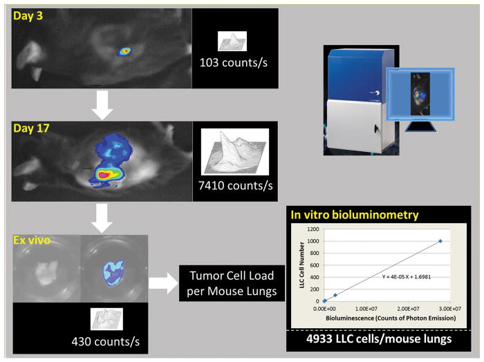
FIGURE 1. Lewis lung carcinoma (LLC) cell-based bioluminescence imaging modality for studying lung cancer cell growth and metastasis.
Shown are images indicative of local growth of LLC cells at days 3 and 17 after subcutaneous injection of LLC cells, and an ex vivo image of the mouse lungs at day 17. Also shown is the LLC cell load in the entire mouse lungs determined by in vitro quantitative bioluminometry in combination with a standard curve of LLC cell number versus bioluminescence intensity.

