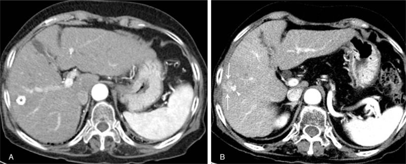
Figure 1.

(A) Contrast-enhanced axial arterial phase CT image shows a compact lipiodolized lesion (asterisk) treated with TACE for a small HCC in the right hepatic lobe. (B) Fourteen-month follow-up contrast-enhanced axial arterial phase CT image depicts arterial enhancement in a recurrent viable HCC (arrows) around a contracted lipiodolized lesion (arrowhead) in the right hepatic lobe. CT = computed tomography, HCC = hepatocellular carcinoma, TACE = transarterial chemoembolization.
