Figure 1.

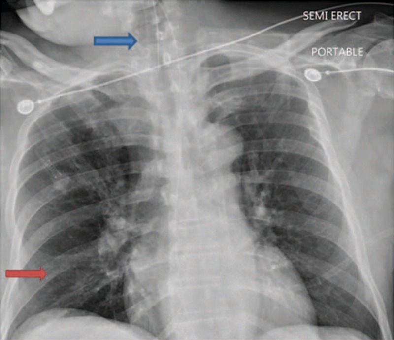
Chest roentgenogram on admission showing ETT high in the trachea (blue arrow), ill-defined infiltrates in right lower lobe (red arrow), and possible infiltrates in the right upper lobe.

Chest roentgenogram on admission showing ETT high in the trachea (blue arrow), ill-defined infiltrates in right lower lobe (red arrow), and possible infiltrates in the right upper lobe.