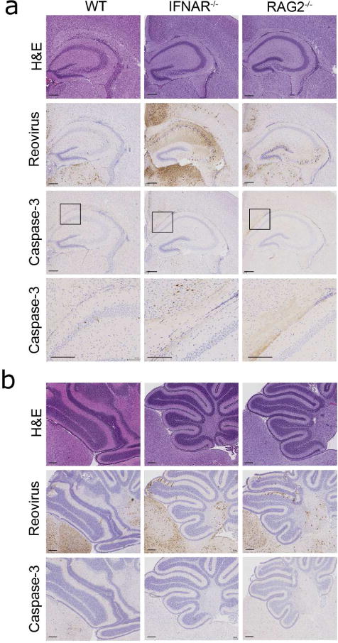
Figure 5. Reovirus tropism in brains of WT, IFNAR−/−, and RAG2−/− mice.

WT, IFNAR−/−, and RAG2−/− mice were inoculated intracranially with reovirus T3D at 100 PFU/g at 4 d of age. At 7 d p.i., brains were removed, the left hemispheres were homogenized for determination of viral titer, and the right hemispheres were processed for immunohistochemistry. Consecutive coronal sections of the brain were stained with H&E, reovirus antiserum, or an anti-cleaved caspase-3 antibody. Representative sections of brains are shown. (a) Hippocampal region stained with H&E or polyclonal reovirus antiserum and higher magnification image of boxed inset stained for cleaved caspase-3 (scale bars, 200 μm). (b) Cerebellum and hind brain. Sections shown are from a WT, IFNAR−/−, and a RAG2−/− mouse with a viral load of 3.9 × 10⁸ PFU/brain, 3.8 × 10⁹ PFU/brain, and 1.2 × 10⁹ PFU/brain, respectively (scale bars, 200 μm).
