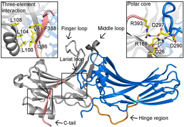
Figure 1. The crystal structure of arrestin-2 in the basal state.

Arrestin-2 (PDB ID: 1G4M) contains two domains, the N- (colored grey) and C-domain (colored blue), which are linked by a hinge region (colored orange). The C-tail (colored pink) of arrestin-2 is attached to the N-domain. One inter-domain contact is the hydrophobic interaction involving β-strand I and the α-helix of the N-domain and the C-tail. It is termed “three-element interaction” and is shown in detail in the upper left box. Another major interaction that holds arrestin-2 in the basal state is the “polar core”, which consists of five charged residues from both domains and the C-tail. It is shown in detail in the upper right box. All participating residues are shown as yellow stick models.
