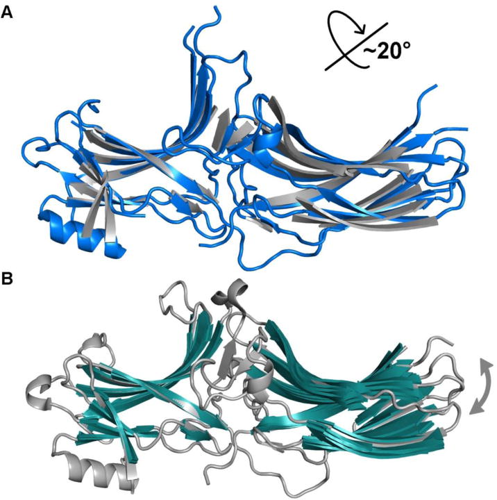
Figure 2. The global conformational change of arrestin upon activation.

A. Superposition of the N-domain of active (colored blue, PDB entry 4JQI) and basal (colored grey, PDB entries 1G4M) arrestin-2 highlights the 20° inter-domain rotation. B. Superposition of the N-domain of basal arrestin-2 variants (colored grey and green, PDB entries 1G4M, 1G4R, 1ZSH, 1JSY, 2WTR, 3GD1, 3GC3) reveals a narrower range of rotational movements between domains in the various basal states.
