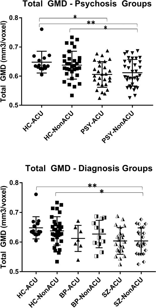
Figure 1. Whole brain gray matter density estimates categorized by the Psychosis (a) or Diagnosis (b) approaches, stratified by adolescent cannabis use.

HC: healthy control; PSY: volunteers with psychosis; SZ: volunteers with schizophrenia; BP: volunteers with psychotic bipolar I disorder; ACU: adolescent onset cannabis use; GMD: gray matter density.
The bar graphs indicate group means, the error bars show standard deviations for total gray matter density estimates.
* p<0.05; ** p<0.01.
